
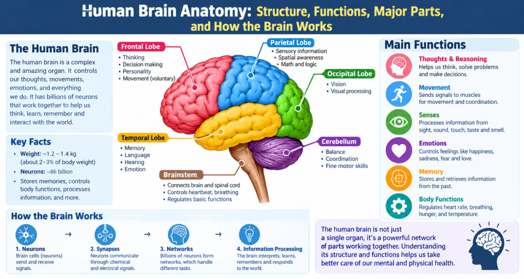

Human brain anatomy is the study of the brain’s structure, major regions, protective layers, and the way different parts work together. The brain is one of the most complex organs in the human body. It helps us think, learn, remember, move, communicate, understand emotions, and respond to the world around us.
Although the brain weighs only around 1.3 to 1.4 kilograms in most adults, it contains billions of nerve cells called neurons. These cells communicate through electrical and chemical signals, creating an enormous network of connections. In simple terms, the brain works like a highly organized communication center, but it’s much more complicated than any computer.
Understanding human brain anatomy can make many everyday processes easier to understand. For example, when you read a sentence, several brain regions work together to recognize letters, understand language, remember meanings, and control eye movements. Likewise, when you decide to pick up a cup, the brain plans the movement and sends signals through the nervous system to the muscles in your hand.
The basic structure of the human brain
The brain is part of the central nervous system, which also includes the spinal cord. It sits inside the skull, where it receives strong physical protection from the bones of the cranium.
At a broad level, the brain can be divided into several major areas:
- Cerebrum
- Cerebellum
- Brainstem
- Thalamus
- Hypothalamus
- Limbic system
- Basal ganglia
- Ventricular system
Each region has specialized roles, but the brain doesn’t work as a collection of isolated parts. Instead, different areas constantly communicate with one another.
For instance, seeing an object involves visual processing areas, attention systems, memory networks, and regions involved in decision-making. This teamwork is one of the most fascinating features of human brain anatomy.
The cerebrum and its important functions
The cerebrum is the largest part of the human brain. It makes up most of the brain’s visible surface and is responsible for many higher mental functions.
The outer layer of the cerebrum is called the cerebral cortex. It’s made mostly of gray matter and contains many nerve cell bodies. Beneath the cortex is white matter, which contains bundles of nerve fibers that connect different areas.
The cerebrum is divided into two major halves, known as the left and right cerebral hemispheres. They are connected by a large bundle of nerve fibers called the corpus callosum.
The two hemispheres communicate continuously. While certain functions are more strongly associated with one side, it would be misleading to say that one hemisphere works completely independently from the other.
Left and right hemispheres
The left hemisphere is often strongly involved in areas such as:
- Language processing
- Certain aspects of reading and writing
- Analytical processing
- Sequential information processing
The right hemisphere contributes significantly to:
- Spatial processing
- Recognition of visual patterns
- Some aspects of attention
- Processing certain forms of nonverbal information
However, most complex activities involve both sides. In everyday life, the hemispheres work together rather than operating as two separate brains.
The four major lobes of the brain
One of the easiest ways to understand human brain anatomy is to learn about the four major lobes of the cerebral cortex.
These lobes are named according to the skull bones located around them:
| Brain lobe | General location | Major functions |
| Frontal lobe | Front of the brain | Planning, decision-making, movement, speech production |
| Parietal lobe | Upper rear area | Touch, body awareness, spatial processing |
| Temporal lobe | Side of the brain | Hearing, memory, language understanding |
| Occipital lobe | Back of the brain | Visual processing |
Although these categories are useful for learning, brain functions don’t always stay inside neat boundaries. Different regions communicate through complex networks.
Frontal lobe
The frontal lobe is involved in planning, reasoning, voluntary movement, attention, and aspects of personality and behavior.
A region called the primary motor cortex helps control voluntary movements. It sends signals to muscles through pathways that eventually travel through the spinal cord and peripheral nerves.
The frontal lobe also contains important language-related areas. In many people, a region commonly called Broca’s area plays an important role in producing spoken language.
Parietal lobe
The parietal lobe processes information related to touch and body position. It also contributes to spatial awareness.
When you touch a warm object, information from sensory receptors travels through nerves toward the spinal cord and brain. The brain then processes the signals so you can identify where the sensation is coming from and what it feels like.
Temporal lobe
The temporal lobes are located on the sides of the brain. They play important roles in hearing, memory, language comprehension, and recognizing objects and faces.
The hippocampus, which is strongly associated with memory formation, is located within the medial temporal lobe.
Occipital lobe
The occipital lobe is positioned toward the back of the brain and plays a major role in vision.
Interestingly, the eyes don’t simply “show” images directly to the brain. Visual information is converted into nerve signals and processed through several stages before the brain creates our conscious visual experience.
The cerebral cortex and gray matter
The cerebral cortex is the thin outer layer covering much of the cerebrum. Its many folds allow a large amount of neural tissue to fit inside the limited space of the skull.
The raised folds are called gyri, while the grooves between them are called sulci. Deeper grooves are sometimes called fissures.
This folded structure increases the surface area of the cortex. In other words, the brain can pack a great deal of processing tissue into a relatively compact space.
Gray matter primarily contains neuron cell bodies and other supporting structures. White matter, located deeper inside the brain, contains many nerve fibers that help different brain regions communicate.
White matter and communication pathways
White matter is an essential part of human brain anatomy because it connects different areas of the nervous system.
Many white matter fibers are covered by myelin, a fatty substance that helps nerve signals travel efficiently along axons.
Some important communication pathways include:
- Corpus callosum, connecting the two cerebral hemispheres
- Internal capsule, carrying important signals between the cerebral cortex and deeper structures
- Association fibers, connecting regions within the same hemisphere
- Projection fibers, connecting the cortex with deeper brain and spinal structures
Think of these pathways as communication routes. A brain region may perform a specialized task, but it often depends on information arriving from other regions.
The cerebellum and movement control
The cerebellum is located toward the back and lower part of the brain, underneath the occipital lobes.
Despite being smaller than the cerebrum, it contains a very large number of neurons. The cerebellum is especially important for coordinating movement, balance, posture, and motor learning.
For example, when someone learns to ride a bicycle, the cerebellum helps fine-tune movements and maintain balance.
The cerebellum doesn’t simply make muscles move. Instead, it helps the nervous system coordinate movements so they become smoother and more accurate.
It also contributes to certain cognitive functions, showing once again that brain regions can have several interconnected roles.
The brainstem and vital body functions
The brainstem connects the brain with the spinal cord. It consists of three main sections:
- Midbrain
- Pons
- Medulla oblongata
The brainstem is essential because it contains pathways that carry information between the brain and spinal cord. It also contains important networks involved in basic life-supporting functions.
The medulla, for example, is involved in regulating functions such as breathing, heart activity, and blood pressure.
The pons contributes to communication between different brain regions and is involved in breathing and sleep-related processes.
The midbrain participates in movement, sensory processing, and reflexes involving vision and hearing.
Because the brainstem contains structures involved in vital functions, damage to this region can have serious consequences.
The thalamus: a major information relay center
The thalamus is located deep within the brain. It acts as an important relay station for information traveling to the cerebral cortex.
Many types of sensory information pass through the thalamus before reaching areas of the cortex responsible for processing them.
The thalamus is also involved in attention, consciousness, sleep, and communication between different brain systems.
There are exceptions to the general sensory relay pattern. For example, much of the sense of smell reaches the cerebral cortex through pathways that don’t follow the same initial thalamic route as other major senses.
The hypothalamus and internal balance
The hypothalamus is a relatively small but extremely important structure located below the thalamus.
It helps maintain the body’s internal balance, a process known as homeostasis.
The hypothalamus contributes to regulation of:
- Body temperature
- Hunger
- Thirst
- Sleep-wake rhythms
- Hormone control
- Stress responses
- Water balance
It also communicates closely with the pituitary gland, an important endocrine gland.
For example, when the body needs to maintain a stable temperature, the hypothalamus helps coordinate responses that support temperature regulation.
The limbic system, emotions, and memory
The term limbic system is commonly used to describe a group of interconnected brain structures associated with emotion, motivation, learning, and memory.
Important structures include the:
- Hippocampus
- Amygdala
- Hypothalamus
- Cingulate cortex and related regions
Hippocampus
The hippocampus plays a major role in forming and organizing certain types of long-term memories.
It’s especially important for learning new information and remembering events and places.
Amygdala
The amygdala is involved in processing emotional information, especially signals related to threat, fear, and emotional significance.
However, emotions aren’t produced by one structure alone. They emerge from interactions among many brain systems.
The basal ganglia and movement
The basal ganglia are groups of structures located deep within the cerebral hemispheres.
They participate in movement control, motor learning, habits, and several cognitive processes.
Rather than directly commanding every movement, the basal ganglia help select and regulate patterns of activity.
This system works closely with the cerebral cortex and cerebellum. When everything is functioning normally, these networks help movements become controlled and coordinated.
Problems involving basal ganglia circuits can contribute to movement disorders, demonstrating how closely brain anatomy and physical function are connected.
The protective layers around the brain
The brain isn’t directly exposed inside the skull. It has several protective systems.
The meninges are three layers of tissue surrounding the brain and spinal cord:
- Dura mater
- Arachnoid mater
- Pia mater
The dura mater is the tough outer layer. Beneath it is the arachnoid mater, followed by the pia mater, which closely follows the surface of the brain.
Between the arachnoid and pia mater is the subarachnoid space, which contains cerebrospinal fluid and important blood vessels.
These structures provide physical protection and support for the nervous system.
Cerebrospinal fluid and the ventricles
The brain contains interconnected spaces called ventricles. These spaces are filled with cerebrospinal fluid, commonly called CSF.
The main ventricles include:
- Two lateral ventricles
- Third ventricle
- Fourth ventricle
CSF circulates through the ventricular system and around the brain and spinal cord.
It helps cushion the nervous system, supports the brain’s chemical environment, and contributes to the removal and movement of certain substances.
The production and circulation of CSF are carefully regulated. When this system becomes disrupted, pressure inside the skull can increase, which may require medical attention.
Blood supply of the human brain
The brain requires a constant supply of oxygen and nutrients. Even though it represents a relatively small portion of total body weight, it uses a significant amount of the body’s energy.
Blood reaches the brain primarily through the:
- Internal carotid arteries
- Vertebral arteries
These vessels contribute to a connected arterial network called the Circle of Willis.
Blood then travels through smaller arteries, arterioles, and capillaries that supply brain tissue.
The brain also has a specialized protective system called the blood-brain barrier. It helps regulate which substances can move from the bloodstream into nervous tissue.
This barrier is extremely important because neurons require a stable chemical environment.
Neurons and glial cells
No discussion of human brain anatomy would be complete without mentioning the cells that make brain activity possible.
Neurons
Neurons are specialized cells that communicate through electrical and chemical signals.
A typical neuron contains:
- Cell body
- Dendrites
- Axon
- Axon terminals
Dendrites often receive information, while the axon carries signals away from the cell body. At connections called synapses, neurons communicate with other neurons or target cells.
Glial cells
The brain also contains many types of glial cells. They support, protect, nourish, and regulate neurons.
Important glial cell types include:
- Astrocytes
- Oligodendrocytes
- Microglia
- Ependymal cells
Glial cells aren’t simply passive support cells. Modern neuroscience shows that they play active and complex roles in brain function.
How different brain regions work together
One of the most important lessons in human brain anatomy is that the brain operates as an interconnected network.
Imagine you’re reading a book. Your visual system processes the letters and words. Language networks help you understand their meaning. Memory systems connect the information to what you already know. Attention systems help you stay focused, while motor systems control your eyes as they move across the page.
At the same time, the brain continues managing breathing, heartbeat, posture, and countless other processes without requiring conscious attention.
This incredible coordination happens continuously.
How the brain controls movement
Voluntary movement begins with activity in brain networks involved in planning and motor control.
A simplified pathway looks like this:
Planning → Motor cortex → Brainstem/spinal pathways → Peripheral nerves → Muscles
However, the actual process is more complicated.
The basal ganglia help select and regulate movement, while the cerebellum contributes to coordination and timing. Sensory information also travels back toward the brain, allowing the nervous system to adjust movement.
That’s why touching an object, reaching for it, and holding it require much more neural processing than they may seem to.
How the brain processes sensory information
Humans receive information through several sensory systems, including:
- Vision
- Hearing
- Touch
- Taste
- Smell
- Balance
- Body position
Specialized receptors detect changes in the environment or inside the body. These signals travel through neural pathways toward the central nervous system.
The brain then processes and combines this information.
For example, when you walk outside, your brain simultaneously processes light, sounds, temperature, balance, body position, and movement. You don’t normally notice each separate process because the nervous system combines them into a continuous experience.
Human brain anatomy and learning
Learning causes changes in the nervous system. When we practice a skill or study new information, patterns of communication between neurons can become stronger or more efficient.
This ability of the nervous system to change is known as neuroplasticity.
Neuroplasticity doesn’t mean the brain can change without limits. Instead, it means the nervous system can adapt in response to experience, learning, development, and certain forms of recovery after injury.
Several factors support healthy brain function and learning, including:
- Adequate sleep
- Regular physical activity
- Balanced nutrition
- Mental stimulation
- Social interaction
- Stress management
- Continued learning
These factors don’t guarantee perfect brain health, but they can support overall well-being.
Common misconceptions about brain anatom
There are plenty of myths about the brain, and some sound convincing at first.
Humans use only a small part of the brain
The popular claim that people use only 10% of their brains is incorrect. Modern neuroscience shows that many brain areas are active across normal daily activities, although activity varies depending on what we’re doing.
The left brain is logical and the right brain is creative
The two hemispheres do have functional differences, but creativity and logical thinking aren’t restricted to one side. Complex abilities usually involve networks across both hemispheres.
Bigger brains always mean greater intelligence
Brain size alone doesn’t determine intelligence or cognitive ability. Brain organization, connectivity, development, genetics, experience, and many other factors matter.
Why understanding human brain anatomy matters
Learning human brain anatomy isn’t only useful for medical students or scientists. It can help anyone understand how the body works.
Knowledge of brain structure is important in fields such as:
- Medicine
- Neurology
- Psychology
- Neuroscience
- Education
- Rehabilitation
- Physical therapy
- Speech and language therapy
- Biomedical research
It also helps people understand why brain injuries, neurological disorders, and developmental conditions can affect movement, memory, communication, or behavior.
Of course, individual medical conditions are complex, and anatomical information alone can’t replace professional medical evaluation.
A simple overview of major brain structures
| Structure | Main role |
| Cerebrum | Thinking, perception, language, voluntary action |
| Cerebral cortex | Higher-level processing |
| Frontal lobe | Planning, movement, decision-making |
| Parietal lobe | Touch and spatial processing |
| Temporal lobe | Hearing, memory, language |
| Occipital lobe | Vision |
| Cerebellum | Coordination and balance |
| Brainstem | Vital functions and communication pathways |
| Thalamus | Major sensory and information relay |
| Hypothalamus | Homeostasis and hormone regulation |
| Hippocampus | Memory formation |
| Amygdala | Emotional processing |
| Basal ganglia | Movement and habit-related circuits |
| Corpus callosum | Communication between hemispheres |
| Ventricles | CSF-filled spaces |
Frequently asked questions about human brain anatomy
What is the largest part of the human brain?
The cerebrum is the largest part of the brain. It contains the cerebral cortex and many deeper structures involved in thinking, perception, movement, language, memory, and other functions.
What are the four main lobes of the brain?
The four major cerebral lobes are the frontal, parietal, temporal, and occipital lobes. Each has specialized functions, although most complex activities involve several regions working together.
What does the cerebellum do?
The cerebellum helps coordinate movement, balance, posture, timing, and motor learning. It works closely with other parts of the nervous system to make movements smoother and more accurate.
What protects the brain?
The skull provides strong physical protection. The meninges, cerebrospinal fluid, and blood-brain barrier provide additional forms of protection and regulation.
What is gray matter?
Gray matter contains many neuron cell bodies and other cellular structures. Much of it is found in the cerebral cortex, but gray matter also exists in deeper brain structures.
What is white matter?
White matter contains many nerve fibers, especially myelinated axons. These fibers connect different brain regions and help information travel efficiently through the nervous system.
Why is the brain divided into two hemispheres?
The two cerebral hemispheres are connected by the corpus callosum and communicate constantly. Although some functions show hemispheric specialization, most complex activities involve cooperation between both sides.
Can the brain change?
Yes. The nervous system has a capacity for change known as neuroplasticity. Learning, experience, development, and some forms of rehabilitation can alter neural connections and activity.
Conclusion: Understanding human brain anatomy
Human brain anatomy reveals an extraordinary biological system made from interconnected regions, cells, blood vessels, protective structures, and communication pathways. From the cerebral cortex and its four major lobes to the cerebellum, brainstem, thalamus, hypothalamus, hippocampus, and basal ganglia, every major structure contributes to the brain’s remarkable ability to coordinate the body and interact with the world.
What’s especially fascinating is that no single brain region works completely alone. Networks communicate continuously, allowing us to learn, remember, move, feel, speak, solve problems, and adapt to new experiences. As neuroscience continues to develop, researchers are discovering more about these connections and the complex organization behind human behavior and cognition.
Stay connected with VoxTrend for the latest ideas, trends, updates & inspiring stories!